In The Diagram Where Is The Mastoid Process
The mastoid process provides an attachment for certain muscles of the neck. The superior border of the mastoid portion of the temporal bone articulates with the parietal bone.
 Mastoiditis
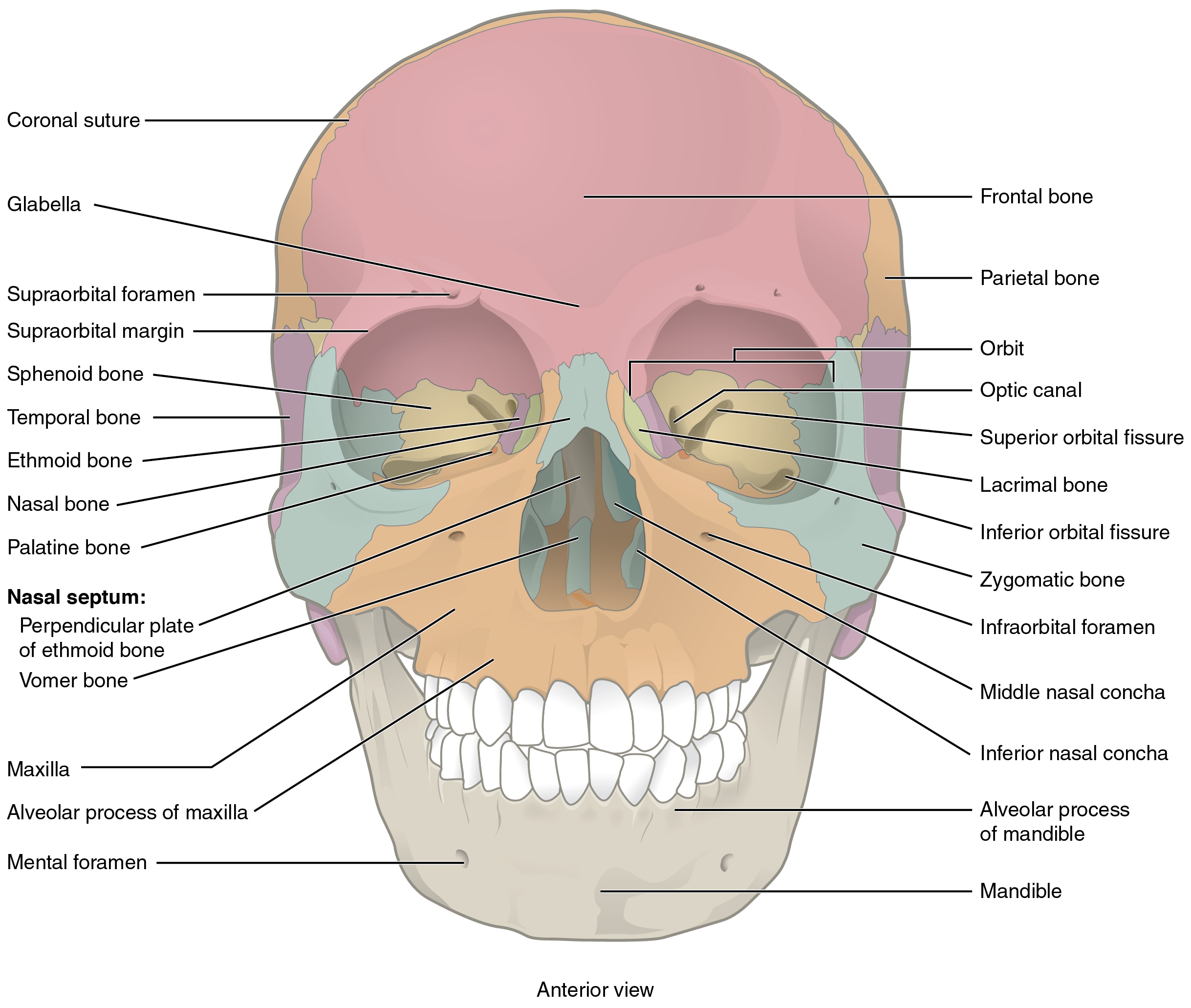
Parts of the maxilla lacrimal ethmoid and sphenoid bones form the medial wall of the orbit.
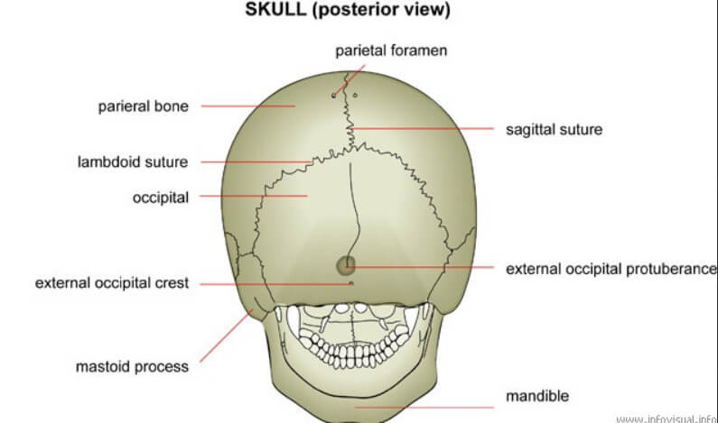
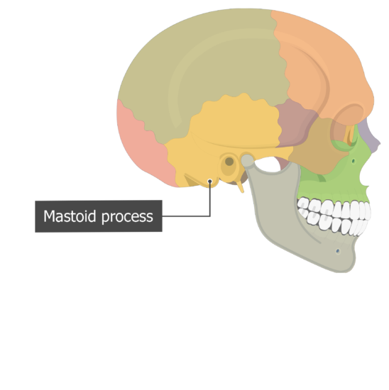
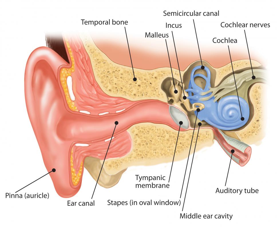
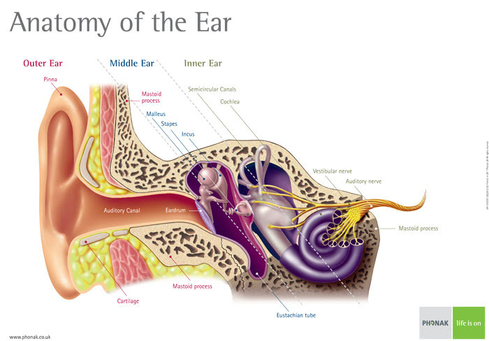
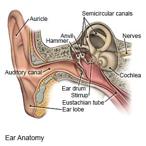
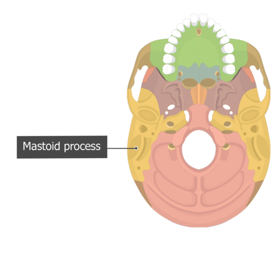
In the diagram where is the mastoid process. It is one of the two projections situated behind the ear. The mastoid process is a pyramidal bony projection from the posterior section of the temporal bone. It is also filled with sinuses or mastoid cells.
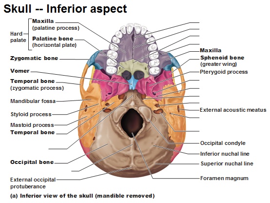
Styloid process pole like process extending downward from the temporal bone on each side of the skull. Parts of the maxilla zygomatic and palatine bones make up the floor of the orbit. Ear infections linked to mastoid cells is typically treated with antibiotics.
Structure and landmarks of the temporal bone. The mastoid process is part of the temporal bone the large bone that runs along the middle bottom of the skull. The mastoid process is located just behind the ear in humans.
It might be a good idea to learn the full anatomy of the skull before zoning in on specific structures like the mastoid practice. The mastoid process is a point of attachment for the sternocleidomastoid muscles of the neck. This article covers the anatomy of the temporal bone its parts connecting sutures and foramina.
Attachment point for muscles and ligaments of neck carotid foramen carotid canal. Mastoid process origin it normally patienthelp. This article covers the anatomy function muscle attachments and clinical aspects of the mastoid process.
Click now to learn more at kenhub. Parts of the frontal and sphenoid bones comprise the roof of the orbit. Mastoid process definition it is a conical or pyramidal bone projection at the base of the skull on each side of the head.
Parts of the zygomatic and sphenoid bones form the lateral wall of the orbit. The mastoid process is located in the posterior portion of the temporal bone. The mastoid process is located just behind the ear canal and lateral to the styloid process.
The mastoid process serves for the attachment of the sternocleidomastoid the posterior belly of the digastric muscle splenius capitis and longissimus capitis.
 Topic 5 Bone Of Skull Neck
 Ce4rt X Ray Positioning Of The Mastoid Process For Radiologic Techs
 Mastoid Process Function Mastoid Process Function Compact Cortical
 Info Body Parts
 Mastoid Process Definition Location Function And Pain
 Mastoid Bone Picture Mastoid Bone Picture Netter 009 Head Neck
 Frontal Bone Parietal Bone Coronal Suture Sphenoid Bone Temporal
 Mastoid Process Poster To Eam And Insertion Point Ecmuscle The
 7 2 The Skull Anatomy And Physiology
 Skull Anatomical Illustrations
 Mastoid Process And Its Relationship With The Middle Ear Photo
![]() Mastoid Process Anatomy Function Attachments Kenhub
Mastoid Process Anatomy Function Attachments Kenhub
 Axial Skeleton Skull
 Axial Skeleton Skull Bones Of The Cranium Bones Of The Face
 Mastoid Muscle For Your Health Muscle Diagram Skeletal Muscle
 Temporal Bone Anatomy
 Duke Anatomy Lab 19 Skull
 Geography Of The Skull
 Temporal Styloid Process Wikipedia
 Canal Wall Reconstruction Mastoidectomy Iowa Head And Neck Protocols
 What Is The Mastoid Process With Pictures
 Mastoiditis What Is It Symptoms Causes Prevention And Treatment
 Cranial Bones Unity Companies Rr School Of Nursing
![]() Mastoid Process Stock Photos Mastoid Process Stock Images Alamy
Mastoid Process Stock Photos Mastoid Process Stock Images Alamy
 Man Dies From Undiagnosed Ear Infection
 Axial Skeleton Skull
 Mastoiditis What You Need To Know
 Styloid Process Strength Of Tears
 Temporal Bone Anatomy
 Solved Axial Skeletona Label The Diagram Of The Skull Anteri
 The Skull Anatomy And Physiology I
 Print Multi Choice The Skeletal System The Axial Skeleton
 Mastoid Process An Overview Sciencedirect Topics
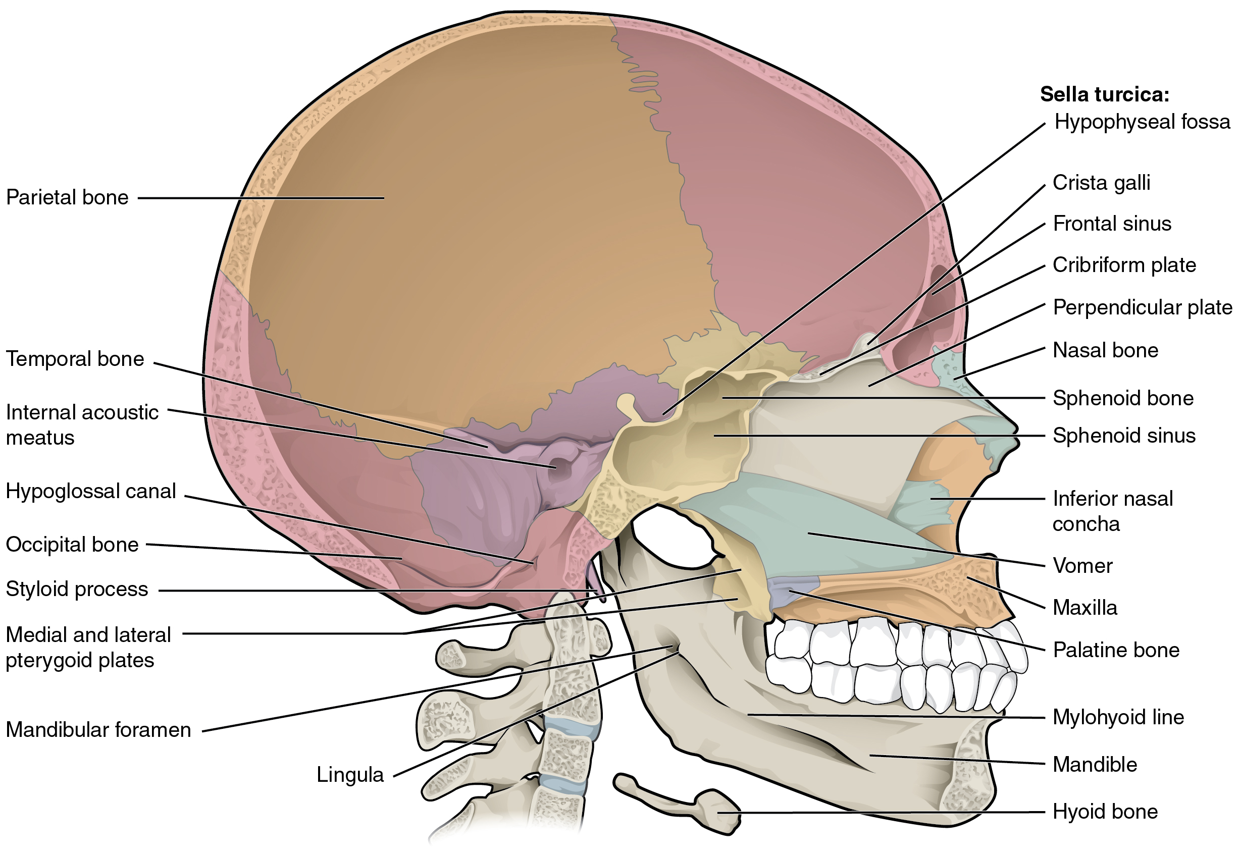 7 2 The Skull Anatomy And Physiology


0 Response to "In The Diagram Where Is The Mastoid Process"
Post a Comment