Nerves Of The Lower Limb Diagram
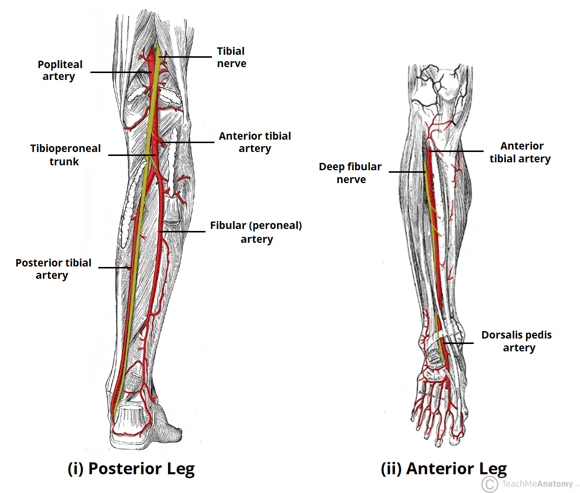
Peripheral nerves and arteries of the lower extremity. The main muscle in this area of the leg is the gastrocnemius which gives the calf a bulging muscular appearance.
 Arteries Of The Lower Limb Thigh Leg Foot Teachmeanatomy
Neurological assessment of the distal lower extremity.

Nerves of the lower limb diagram. Anatomy course function and clinical significance of the sciatic nerve. Atlas main nerves of the lower extremity. Modern texts are in agreement about which areas of the skin are served by which nerves but there are minor variations in some of the details.
The anterior tibial posterior tibial and the fibular arteries supply blood to. Nerves of lower limb diagram 1000 images about the body anatomy help on pinterest leg nerves of lower limb diagram. Nerves of the lower limb.
The lumbar plexus forms in the lower back from the merger of spinal nerves l1 through l4 while the. Some nerves of the sacral plexus innervate this area namely the superficial fibular nerve the deep fibular nerve and the tibial nerve. In this article you will find the anatomy branches and mnemonics related to the lumbar plexus.
Ask the patient to dorsi flex the foot l4 and extend the toes l5. The main artery of the lower limb is the femoral artery and its continuationthe popliteal artery. The femoral artery supplies the gluteal region and the thigh before it continues as the popliteal artery in the posterior knee.
The nerves of the leg and foot arise from spinal nerves connected to the spinal cord in the lower back and pelvis. Back to nerves of lower limb diagram. Major nerves of the hip thigh lower leg and foot.
The superficial fibular nerve. As these nerves descend toward the thighs they form two networks of crossed nerves known as the lumbar plexus and sacral plexus. Ask the patient to evert the foot.
Cutaneous innervation refers to the area of the skin which is supplied by a specific nerve. The borders designated by the diagrams in the 1918 edition. The common fibular nerve.
Cutaneous innervation of the lower limbs. 12 photos of the nerves of lower limb diagram. Sensory as per cutaneous innervation in side diagrams.
Major nerves of the hip thigh lower leg and foot. Nerves of lower limb diagram anatomy nerve supply to the upper limb geeky medics photo nerves of lower limb diagram anatomy nerve supply to the upper limb geeky medics image nerves of lower limb diagram anatomy nerve supply to the upper limb geeky medics gallery. Nerves of lower limb diagram 1000 images about the body anatomy help on pinterest leg.
Arteries veins and nerves of the lower limb a diagram.
 Lumbar Plexus And Derivative Nerves Muscles And Lesions Of The
 Medial Lower Leg Muscles Diagram Vmglobal Co
 Nerve Supply Of The Lower Limb Ppt Video Online Download
 Cutaneous Innervation Of The Lower Limbs Wikipedia
 Nerves Of Body Circular Flow Diagram
 Core Anatomy Lower Limb Dermatomes Frcem Primary Blog
 Prac 6 Nerves Of Lower Limb Diagram Quizlet
 Functional Regional Anesthesia Anatomy Nysora
 Duke Anatomy Lab 14 Anterior Thigh Leg
 Nerves Of The Lower Limb Teachmeanatomy
 Peripheral Nervous System Anatomy Overview Gross Anatomy
 Cutaneous Innervation Of The Lower Limbs Wikipedia
 Anatomy Mbbs Lower Limb Nerves Vessels Lymphatics
 Instant Anatomy Lower Limb Nerves Femoral
 Femoral Nerve Anatomy Orthobullets
 How To Assess Sensation Neurologic Disorders Merck Manuals
 Anatomy Revision Of The Upper Limb Lower Limb Back
 Nerves Of The Lower Limb Diagram Quizlet
 Lower Limb Bones Muscles Joints Nerves How To Relief
 Instant Anatomy Lower Limb Nerves Sciatic Lower Extremity
 Easy Notes On Cutaneous Innervation Of The Lower Limb Earth S Lab
 The Left Panel Shows The Anterior View Of Veins In The Legs And The
 Module 2 Lower Extremity Orthopedic Imaging
 New Photos In Nerves Of The Lower Limb Anatomy
 Lower Extremity Anatomy Bones Muscles Nerves Vessels Kenhub


0 Response to "Nerves Of The Lower Limb Diagram"
Post a Comment