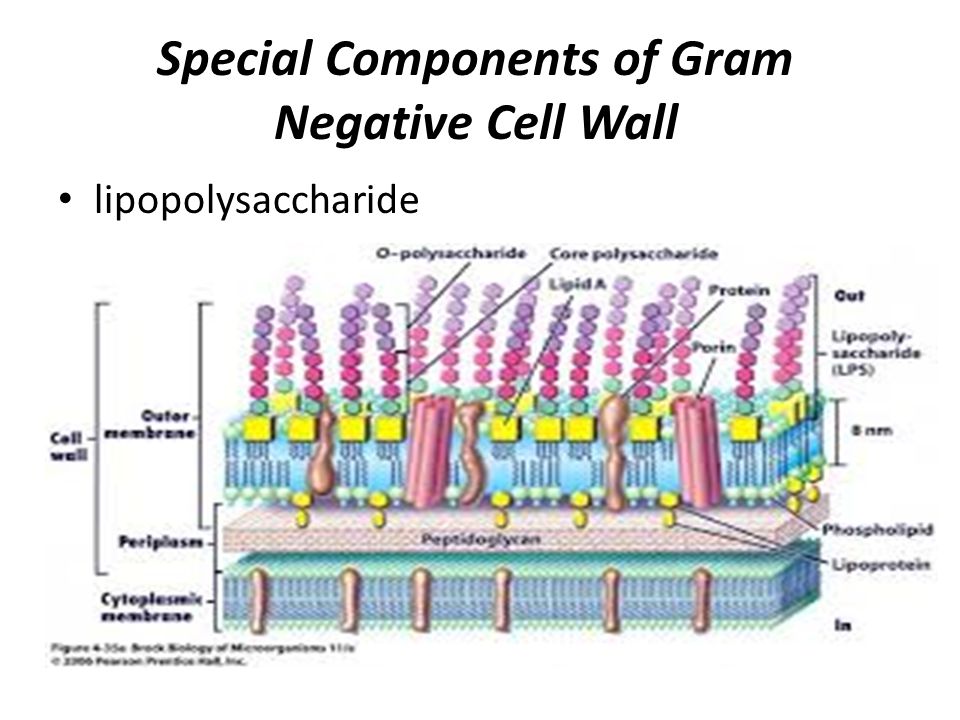
Gram Negative Cell Wall Diagram
800 401 pixels. The cytoplasm is enclosed by three layers the outermost slime or capsule the middle cell wall and inner cell membrane.
 Bacterial Morphology And Structure Ppt Video Online Download
D it contains teichoic acids.

Gram negative cell wall diagram. You can safely assume that the cell. Can be decolorized to accept counterstain safranin and stain pink or red. C it protects the cell in a hypertonic environment.
Retain crystal violet dye and stain blue or purple. Diagram demonstrating of the cell wall structure a grampositive. In gram positive bacteria the cell wall is much thicker 20 to 40 nanometers thick.
Gram negative cell wall structure download full size image cell walls what is the difference between gram positive and negative bacteria. Size of this png preview of this svg file. Cell wall is 8 12 nm thick.
Thick multilayered thin single layered 5. In figure 43 which diagram of a cell wall is a toxic cell wall smaller gram negative in figure 43 which diagram of a cell wall has a wall that protects against osmotic lysis. In peptidoglycan makes up as much as 90 of the thick cell wall enclosing the plasma membrane.
2 each of the following statements concerning the gram positive cell wall is true except a it maintains the shape of the cell. The wall is smooth. Filegram negative cell wallsvg.
25 shows a typical prokaryotic structure. The wall is wavy. This is a file from the wikimedia commons.
Cell wall is 20 30 nm thick. You have isolated a motile gram positive cell with no visible nucleus. Posted on march 29 2019 by admin.
Information from its description page there is shown below. 320 160 pixels 640 320 pixels 1024 513 pixels 1280 641 pixels 1486 744 pixels. On adding a counterstain such as safranin or fuchsine after washing gram negative bacteria are stained red or pink while gram positive bacteria retain their crystal violet dye.
Diagram of gram positive and negative bacteria cell wall. This is due to the difference in the structure of their bacterial cell wall. In this article we will discuss about the cell structure of bacteria with the help of diagrams.
The cell wall of gram negative bacteria is thin approximately only 10 nanometers in thickness and is typically comprised of only two to five layers of peptidoglycan depending on the growth stage. B it is sensitive to lysozyme. The major cytoplasmic contents are nucleoid plasmid.
During these thick multiple layers 2080 nm of peptidoglycan retain the dark purple primary stain crystal violet whereas gram negative bacteria stain pink. A bacterial cell fig. E it is sensitive to penicillin.
 Bacteria Cell Walls Microbiology
 Schematic Structure Of Gram Positive And Gram Negative Cell Walls
 Tackling Multi Drug Resistant The Cell Wall Of Gram Negative
 Cell Wall Structure Gram Negative Bacteria Example Helicobacter
 Cell Wall Of Bacteria Structure Functions Gram Positive And Gram
 Liquid Crystalline Bacterial Outer Membranes Are Critical For
 Cell Wall Of Bacteria Structure Functions Gram Positive And Gram
 Gram Positive Vs Gram Negative Bacteria
 The Outer Membrane Is An Essential Load Bearing Element In Gram
 Step 1 Microbiology Basic Bacteriology Flashcards Memorang
 A Comparison Of The Cell Walls Gram Positive And Gram Negative
 Multiple Choice Quiz On Bacterial Cell Wall Biology Multiple
 Gram Negative Bacteria Wikipedia
 Outer Membrane Proteins Simbac Simulations Of Bacterial Systems
 Cell Wall Definition Structure Function With Diagram Sciencing
 Gram Negative Cell Wall Diagram Amazing Gram Positive And Negative
 Gram Negative Cell Wall Diagram Quizlet
 Lipopolysaccharide Lps Of Gram Negative Bacteria Characteristics
 Bacterial Cell Wall Structure Composition And Types Online
 What Is The Difference Between Gram Positive And Gram Negative
 Bacteria 101 Cell Walls Gram Staining Common Pathogens Tusom
 Bacterial Cell Wall Structure Composition And Types Online


0 Response to "Gram Negative Cell Wall Diagram"
Post a Comment