Identify The Structures Labeled A B And C In The Diagram Of A Sarcomere Above
D after the contraction ends. Identify major abdominal arteries.
 The Sarcomere And Sliding Filaments In Muscular Contraction
Model 3 muscle contraction 13.
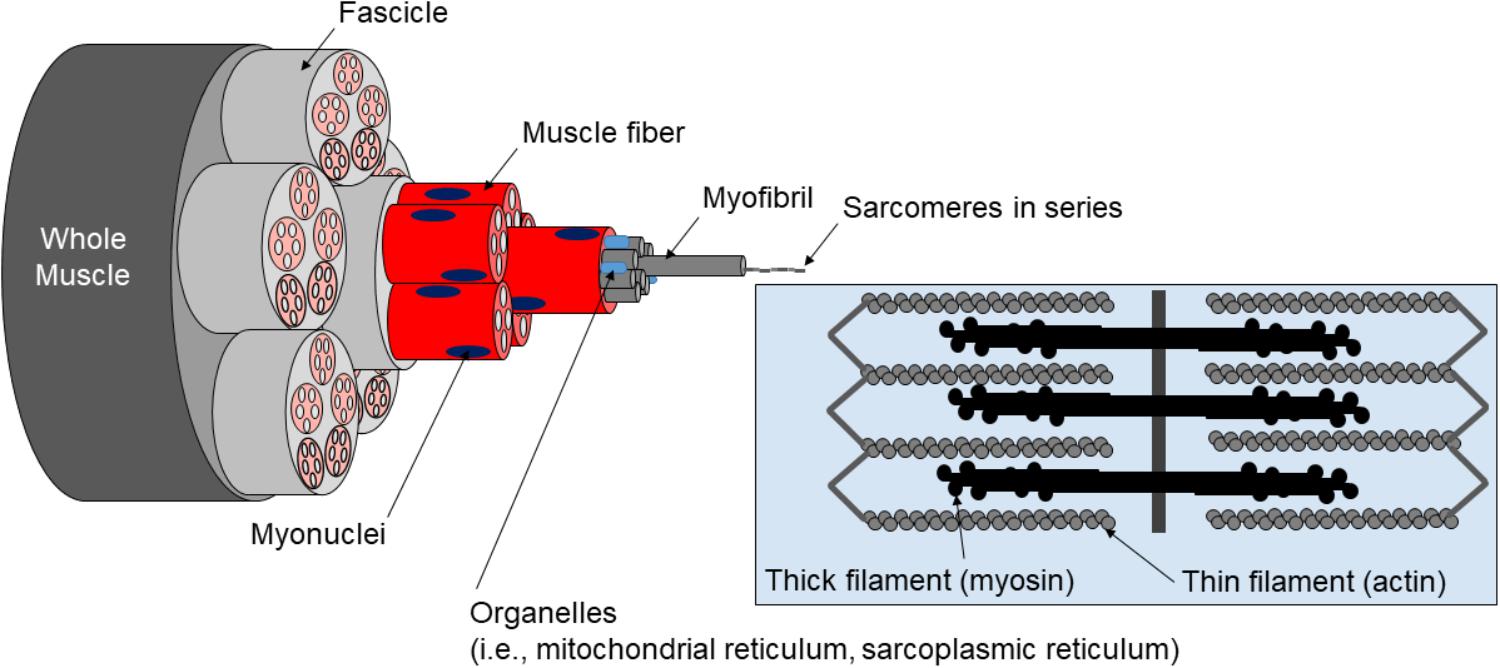
Identify the structures labeled a b and c in the diagram of a sarcomere above. Draw and label a diagram to show the structure of a sarcomere including z lines actin filaments myosin filaments with heads and the resultant light and dark bands. The region at the center of an a band of a sarcomere that is made up of myosin only. Label the thick and thin filaments in figs.
Label each of the lines. B in sarcomere 2 identify the location within the sarcomere of the cross section indicated by figure b in model 3. In the diagram below draw three vertical lines showing the locations within a sarcomere of the cross sections indicated by figures a b and c.
A b c fig. Which of the figures a b or c represents a cross section in the h zone. Draw a vertical line and label it a.
A b and c above. A short l there is overlap of thin filament into the region of the thick filament with no myosin head or cross bridges. E all of these answers are correct.
The diagrams in model 3 are cross sections of a sarcomere that show the filaments at various locations within a sarcomere. Label the thick and thin filaments in figs. Draw a vertical line and label it b.
B in response to acetylcholine binding to ca2 release channels. The h zone gets shorter and may disappear during muscle contraction. A b and c above.
Sarcomere 1 sarcomere 2 sarcomere 3 a in sarcomere 1 identify the location within the sarcomere of the cross section indicated by figure a in model 3. C in sarcomere 3 identify the location within the sarcomere of the cross section indicated by figure c in model 3. Difficulty hard learning objective 1 149 identify the termination of the.
In the diagram where would you find stored ca2 a b b d c g d f e k answer d from anatomy 2085 at florida international university. Draw a vertical line and label it b. One of the components of a myofibril is the structure labeled.
Draw a vertical line and label it a. B in sarcomere 2 identify the location within the sarcomere of the cross section indicated by figure b in model 3. Match the terms to the brain areas.
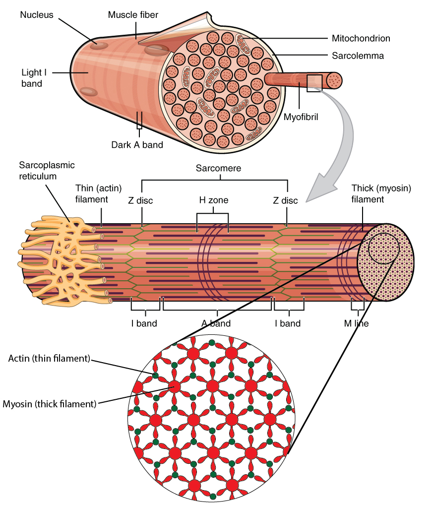
Describe the structure of striated muscle fibers including the myofibrils with light and dark bands mitochondria the sarcoplasmic reticulum nuclei and the sarcolemma. As the length is increased towards lo more and more possible cross bridges may be formed when lo is exceed then there will be mysoin heads in the center of the sarcomere that cant bind to actin sites and. C by active transport using ca2 pumps in the sr membrane.
Which of the figures a b or c represents a cross section in the h zone. In the diagram below draw three vertical lines showing the locations within a. Play this quiz called label the sarcomere and show off your skills.
Label the spinal column. A at the beginning of a contraction. Various locations within a sarcomere.
Calcium ions are released from the sarcoplasmic reticulum into the cytosol. Identify structures of the heart. Draw a vertical line and label it c.
Sarcomere Length Dependent Effects On Ca2 Troponin Regulation In
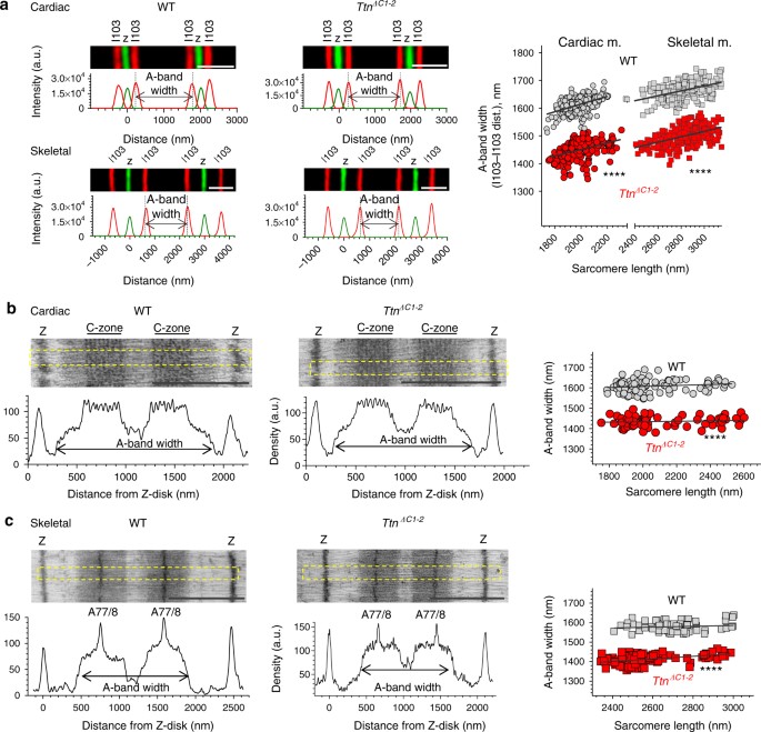 The Giant Protein Titin Regulates The Length Of The Striated Muscle
 Homework 10b Homework 10b Muscle Tissue Sarcomeres And Mechanisms
 Muscle Specific Stress Fibers Give Rise To Sarcomeres In
Biol 237 Class Notes Muscle Cells Muscle Physiology
 Practical Human Anatomy Quiz 091239 Human Pathophysiology
 A P Final Ch 10 At Wayne State University Studyblue
 A Schematic Representation Of A Sarcomere B Model Of The Assembly
 Muscle Specific Stress Fibers Give Rise To Sarcomeres In
10 2 Skeletal Muscle Anatomy Physiology
Sarcomere Length Dependent Effects On Ca2 Troponin Regulation In

11 2 Muscles And Movement Bioninja
 Frontiers A Critical Evaluation Of The Biological Construct
 Inter Sarcomere Coordination In Muscle Revealed Through Individual
 Muscle Specific Stress Fibers Give Rise To Sarcomeres In
 Muscle Tissue Junqueira S Basic Histology Text And Atlas 15e
 Identify The Structures Labeled A B And C In The Diagram Of A


0 Response to "Identify The Structures Labeled A B And C In The Diagram Of A Sarcomere Above"
Post a Comment