Which Protein Subunits Are Depicted In The Diagram
Select all that apply. A single amino acid monomer may also be called a residue indicating a repeating unit of a polymer.
The Lactate Receptor Hcar1 Modulates Neuronal Network Activity
Elf5 gtp is now bound to elf1a in the a site.
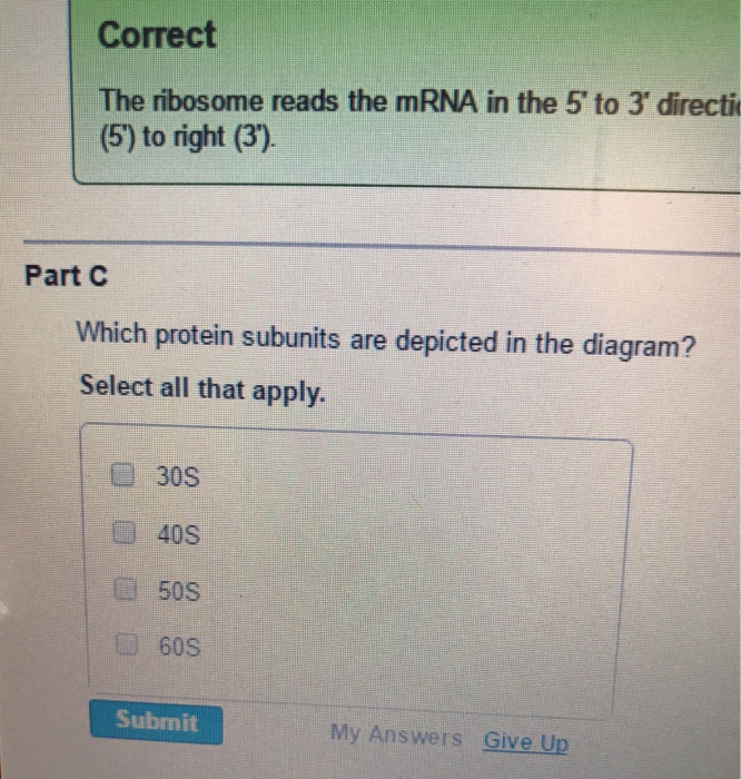
Which protein subunits are depicted in the diagram. Different subunits belonging to the same protein plex different subunits belonging to the same protein plex often exhibit discordant expression levels and evolutionary properties figure s1 a mechanism of covalent substrate binding in the x ray fig 1 structure of the dhakdha plex a ribbon diagram of the dhak dimer the subunits are gray the n terminal. Home study science biology biology questions and answers the ribosome in the diagram is in the process of synthesizing a protein using directions transcribed. Protein what kind of foodwhat s normal protein levelswhich protein powder ketowhich protein.
Part a identify the the following elements on a diagram of translation. 8 correct association of the 40s and 60s subunits induces hydrolysis of the gtp bound to elf5. The ribosome in the diagram is in the process of synthesizing a protein using directions transcri.
A small protuberance called a platform extends from the base. Drag the labels to their appropriate locations in the diagram. This accurately describes the polarity with which the message is read as well as the direction of protein synthesis.
A site is a site where the charged trna enters the complex. Part c which protein subunits are depicted in the diagram. Proteins form by amino acids undergoing condensation reactions in which the.
Also describes functional dissection of the nascent polypeptide associated plex and labeled as. Term ggua my answers give up correct small unit complexes with eukaryotic initiation factor and a charged trnam t nitiation factors disassociate and are replaced ongation factors as the tran ferase a peptide bond bet. It then adds the amino acid specific to the codon sequence of the mrna to the growing polypeptide chain at p site.
Extending from the main body of the large subunit are a stalk and central protuberance. A p and e sites. Correct the subunits present depend on whether the ribosome is eukaryotic 40s and 60s or prokaryotic 30s and 50s.
Association of the subunits to form the 70 s monomer is depicted in figure 22 5b. Which protein subunits are depicted in the diagram entitled as functional dissection of the nascent polypeptide associated plex which protein subunits are depicted in the diagram. Proteins are polymers specifically polypeptides formed from sequences of amino acids the monomers of the polymer.
The ribosome moves down the mrna in the 5 to 3 direction and synthesizes protein in the direction of amino terminus to carboxyl terminus. Ribosomes and protein synthesis. The 50 s subunit is somewhat more spherical and possesses a flattened region on one surface fig.
7 the 60s subunit will now combine with the 40s subunit resulting in release of most of the initiation factors. Protein structure is the three dimensional arrangement of atoms in an amino acid chain molecule. Then elf5b gdp and elf1a are released.
The small and large subunits of the ribosome form the complex around the start codon of the mrna. It created three sites namely.
Expression Purification And Antimicrobial Activity Of S100a12
 The Crystal Structure Of Staufen1 In Complex With A Physiological
 Tnip2 Is A Hub Protein In The Nf Kb Network With Both Protein And
 Frontiers Meloidogyne Incognita Passe Muraille Mipm Gene Encodes
 Amazon Com Cellular Plant And Animal Anatomy Notebook Chart
 The Er Membrane Protein Complex Promotes Biogenesis Of Sterol
113 Gene Expression The Process Of Gene Expression Simply Refers To
Team Unsw Australia Lab Cloning 2018 Igem Org
 Making Life From Scratch American Scientist
Ser Thr Protein Phosphatases In Fungi Structure Regulation And
 Ribbon Diagram An Overview Sciencedirect Topics
Ser Thr Protein Phosphatases In Fungi Structure Regulation And
Components Of The Dcc A E The Protein Subunits Their Domain
The Protein Lipidation And Its Analysis
 3 4 Protein Synthesis Anatomy And Physiology
Structure Of The Human Mitochondrial Ribosome Studied In Situ By
Ch103 Chapter 8 The Major Macromolecules Chemistry
 Figure 8 From Sequential Domain Assembly Of Ribosomal Protein S3
 Binding Assays Of Variant Coat Proteins And Dec To Probe The
 Evolutionary Shift Toward Protein Based Architecture In Trypanosomal
 Reduced Proteasome Activity In The Aging Brain Results In Ribosome
Substrate Assisted Mechanism Of Catalytic Hydrolysis Of
 Solved 7 Hemoglobin Pictured Below And To The Left Con


0 Response to "Which Protein Subunits Are Depicted In The Diagram"
Post a Comment