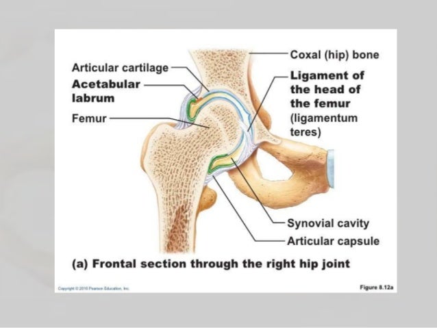
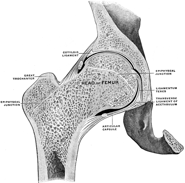
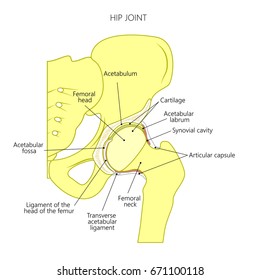
The Diagram Shows A Frontal Section Of The Hip Joint
Ligament of the bead of the femur the shoulder joint is built for mobility. Joint between forearm bones and wrist e.
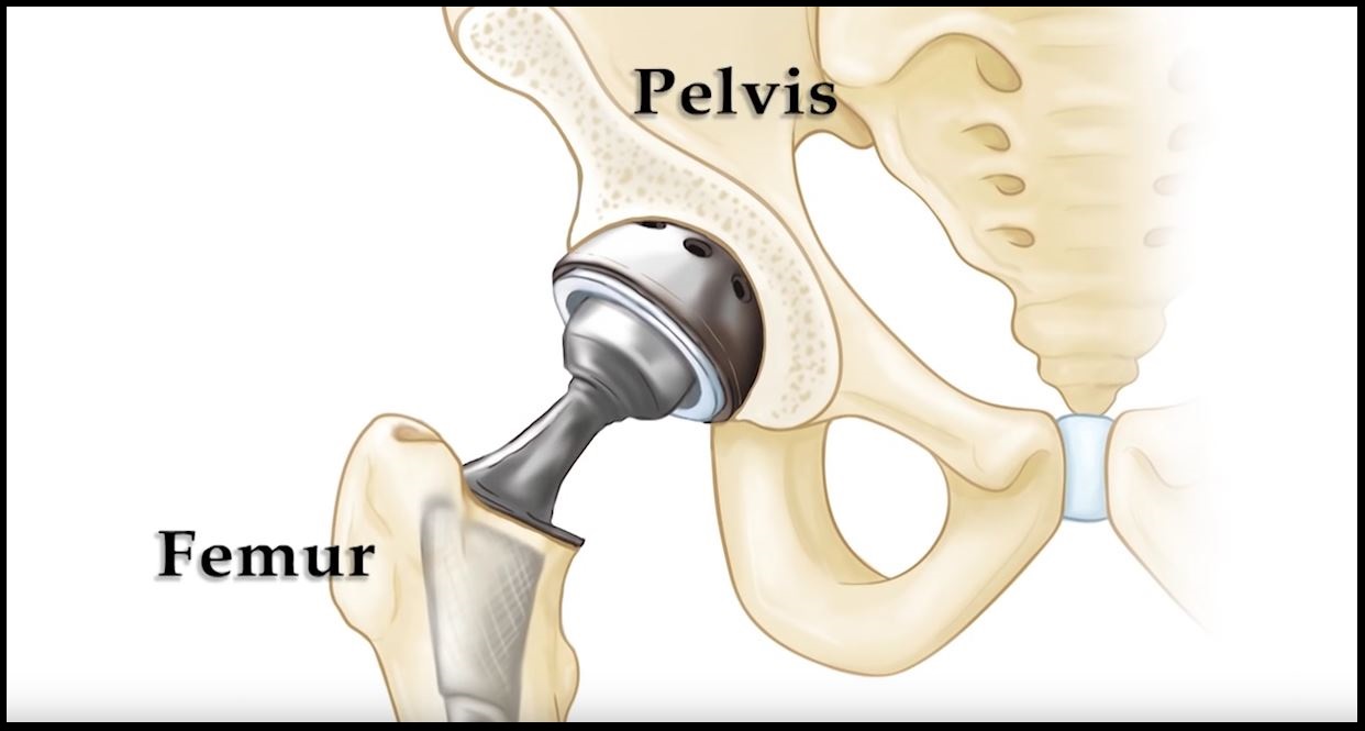 Dislocation After Hip Replacement Orthoinfo Aaos
The hip joint is an example of a synovial joint.

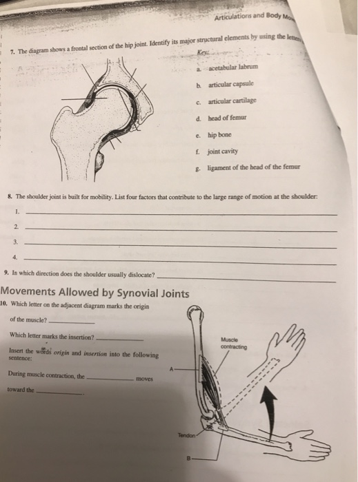
The diagram shows a frontal section of the hip joint. The diagram on the right shows a cross section of the hip. Identity its major structural elements by using the letters key. The diagram shows a frontal section of the hip joint.
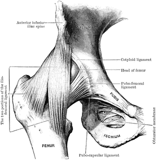
Identify its major structural elements by using the key letters. Intervertebral joints between articular processes d. Hip anatomy function and common problems front view of the hip joint bones normally a smooth cushion of shiny white hyaline or articular cartilage about 14 inch thick covers the femoral head and the acetabulum.
The diagram shows a frontal section of the hip joint. Movement of a limb away from the midline or median plane of the body in the frontal plane is known as. As you can see the top of the femur is shaped like a ball and the concave cavity of the pelvis is shaped like a socket.
Head of femur e. The diagram shows a frontal section of the hip joint. The diagram below shows a frontal section of hip joint posted on december 31 2016 by admin frontal cut view of the hip resurfacing geometrical model to bring you back seventh grade math class the shape above is called an isosceles trapezoid when in central position and it formed by ground the full body weight 52.
Head of femur f. This type of movement is common in ball and socket joints and can be described as the movement of a bone around its longitudinal axis. Ligament of the head of the femur g.
Joint between skull bones a. Ball and socket 2. The diagram on the left is a back view of the hip joint showing the thigh bone femur going into the pelvis bone held together by ligaments.
Joint between the axis and atlas b. A it is the fluid secreted by the knee joint that stabilize the joint bit is the partial fibrous capsule of the knee joint cit is the lateral ligaments of the knee that prevent hyper extension d it is articular cartilage that prevents the knee from rotating. Identify its major structural elements by using the key letters.
Diagram of frontal section the hip joint posted on june 4 2016 by admin neck of femur pelvis greater trochanter diagram hip joint cartilage pubic remus head patient perfect hip joint anatomy 52 for your sacrum and coccyx with frontal section of hip joint awesome ideas 6 image not available the anatomical position or neutral is starting for.
 Joints Ligaments And Connective Tissues Advanced Anatomy 2nd Ed
 Clinical Anatomy Of The Elbow And Shoulder Reumatologia Clinica
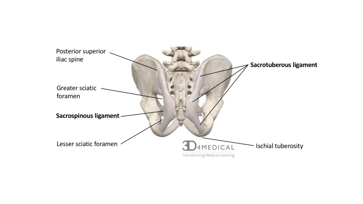
 The Hip Complex
 The Hip Joint Articulations Movements Teachmeanatomy
 Electromyographic Study Of The Hip Abductor Muscles As Subjects With
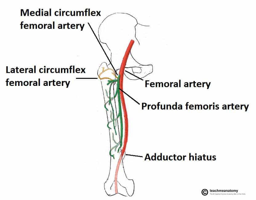
 Anatomy Of Lower Extremity
 The Hip Complex
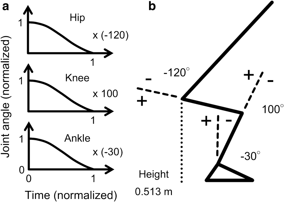 Effect Of Hip Joint Angle At Seat Off On Hip Joint Contact Force
 Hip Abduction Exercises Anatomy Benefits Effectiveness
 Hip Joint Anatomy Pictures And Information
 The Hip Complex Joint Structure And Function A Comprehensive
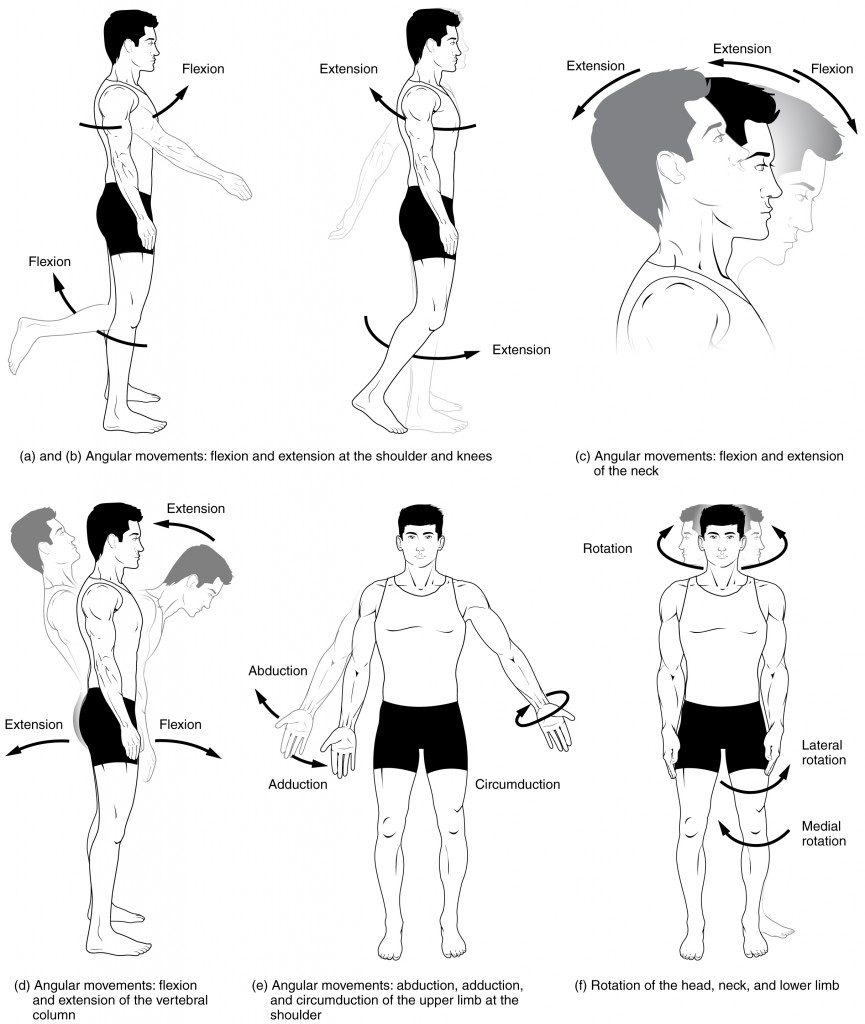 9 5 Types Of Body Movements Anatomy And Physiology
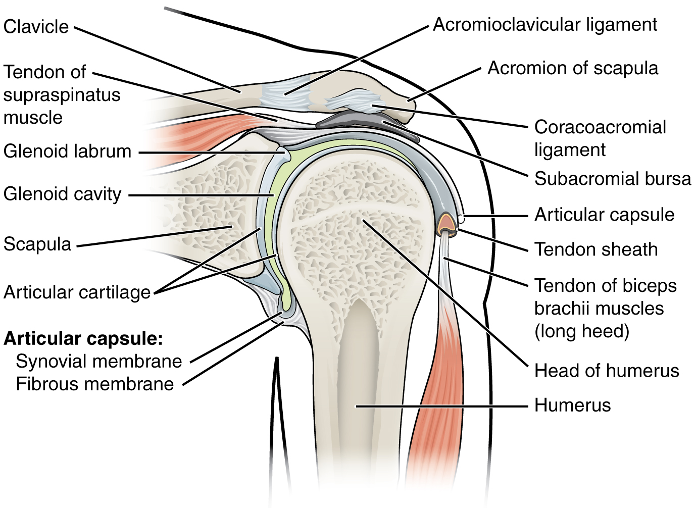
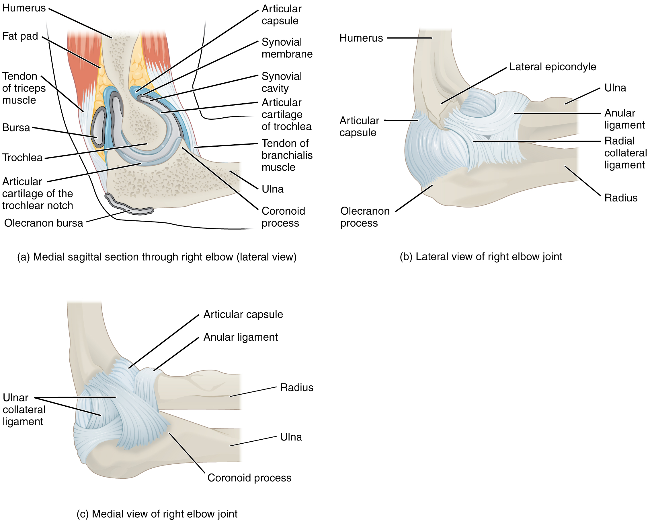 9 6 Anatomy Of Selected Synovial Joints Anatomy And Physiology
Summary Shs111 Shs111 23 Oct 2015 Studocu

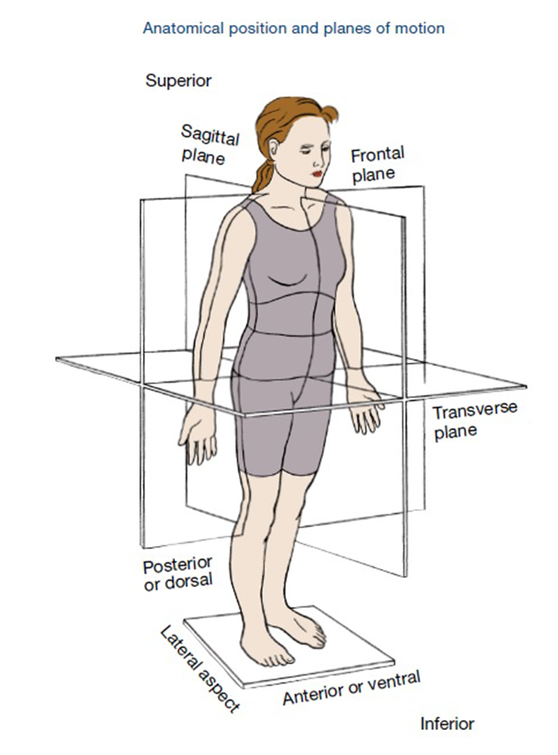 Planes Of Motion Explained Ace Blog
 9 6 Anatomy Of Selected Synovial Joints Anatomy And Physiology
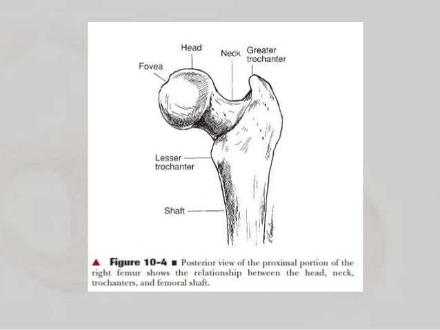
The Femur Human Anatomy
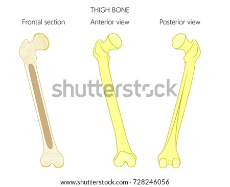 Vector Illustration Anatomy Thigh Bone Tubular Stock Vector Royalty
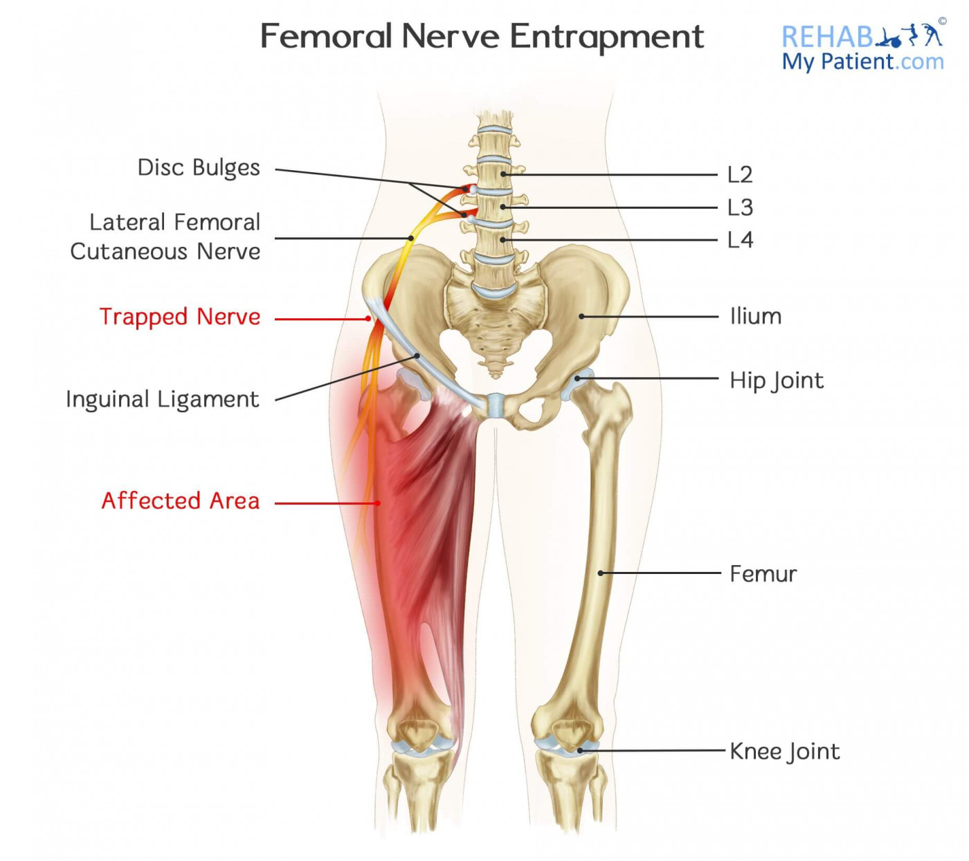 Femoral Nerve Entrapment Rehab My Patient
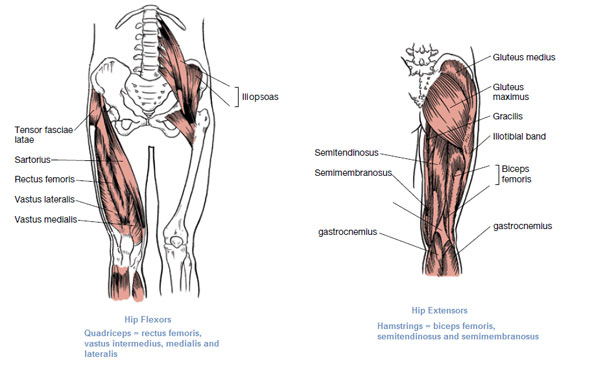
![]() Diagram Pictures Muscles Of The Hip And Thigh Anatomy Kenhub
Diagram Pictures Muscles Of The Hip And Thigh Anatomy Kenhub
 Anatomy Of The Hip Joint Radiology Case Radiopaedia Org
 The Diagram Shows A Frontal Section Of The Hip Joint I Chegg Com
Gcse Pe Structured Questions 1a Applied Anatomy And Physiology
 Pearson Etext15 Review 7 The Diagram Shows A Frontal Section Of
 Classification Of Joints Anatomy And Physiology I
 Muscles That Move The Leg
 Frontal Section Of Hip Joint Clipart Etc
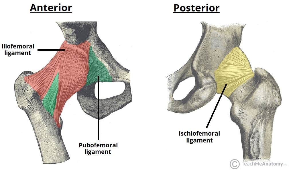 The Hip Joint Articulations Movements Teachmeanatomy
 Labrum Images Stock Photos Vectors Shutterstock
 Femur Wikipedia
0 Response to "The Diagram Shows A Frontal Section Of The Hip Joint"
Post a Comment