In The Figure Which Diagram Of A Cell Wall Is A Gram Negative Cell Wall
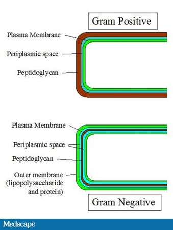
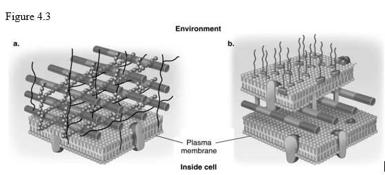
In figure 43 which diagram of a cell wall is decolorized by ethyl alcohol. The cell wall of gram positive bacteria is high in peptidoglycan which is responsible for retaining the crystal violet dye.
 2
2
In figure 43 which diagram of a cell wall has a wall that protects against osmotic lysis.

In the figure which diagram of a cell wall is a gram negative cell wall. In the figure which diagram of a cell wall has a structure that protects against osmotic lysis. In the figure which diagram of a cell wall has a structure that protects against osmotic lysis. This is due to the difference in the structure of their bacterial cell wall.
A a b b c both a and b d neither a nor b e the answer cannot be determined based on the information provided. In figure 43 which diagram of a cell w all possesses molecules responsible for associated with infection. Peptidoglycan pep tid o gly can is a molecule found only in the cell walls of bacteria.
Aspergillus fumigatus mads box transcription factor rlma is required. B the one on the right front 30. In figure 43which diagram of a cell wall is a gram negative cell wall.
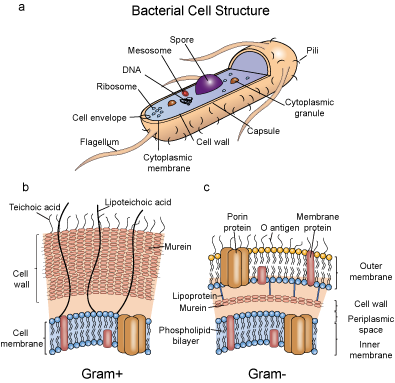
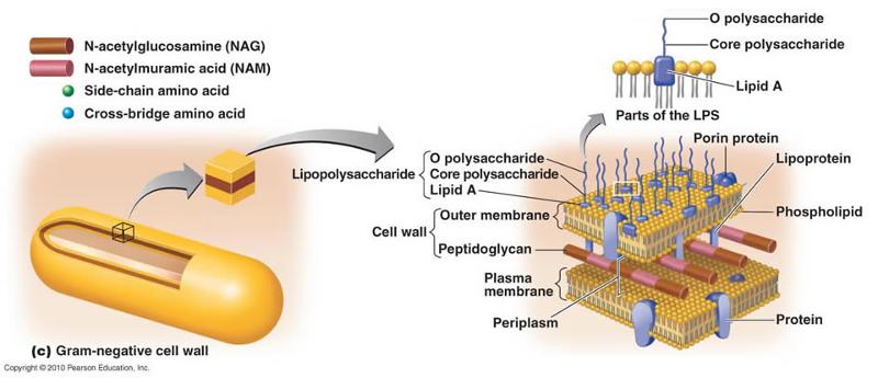
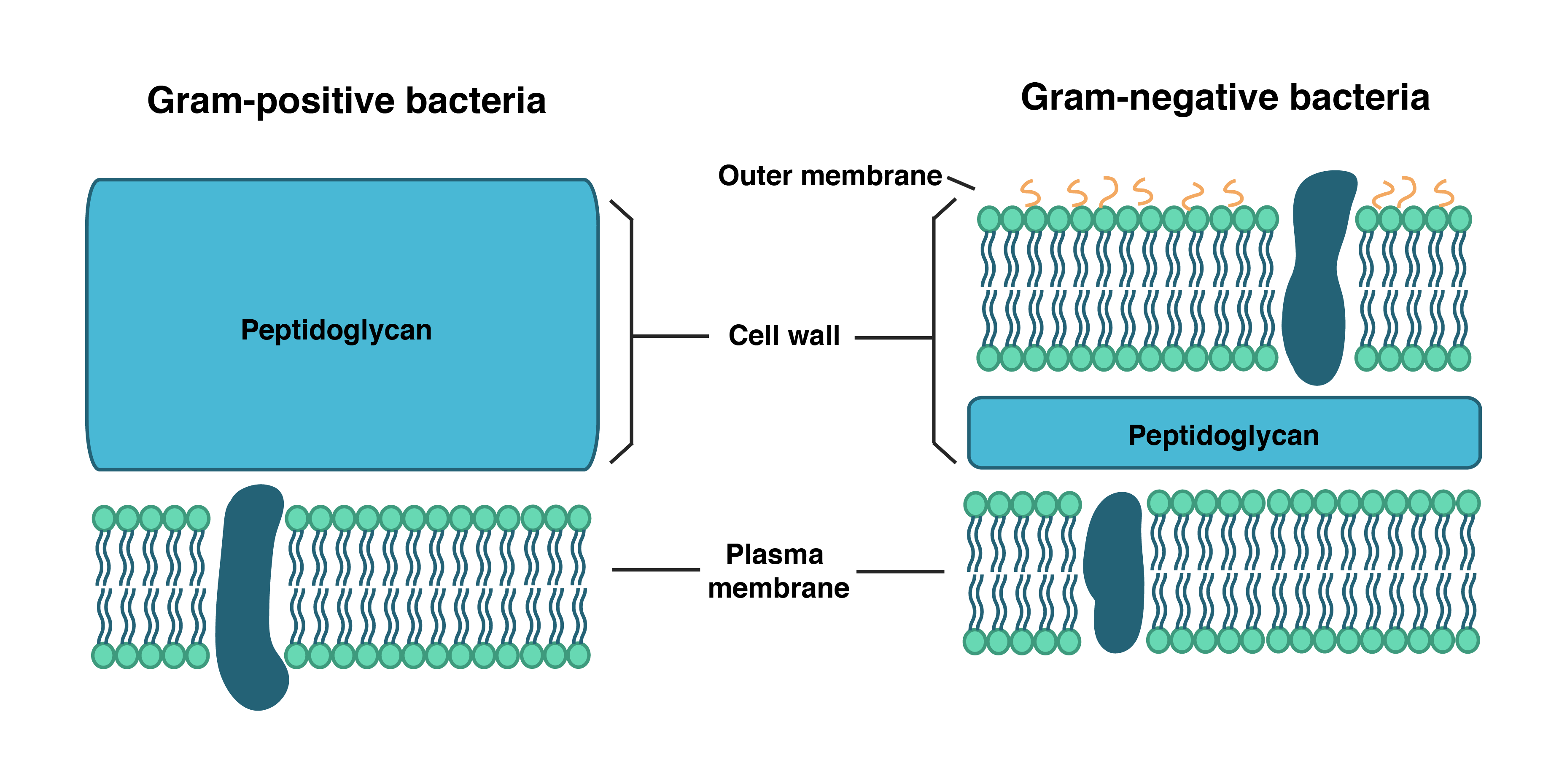
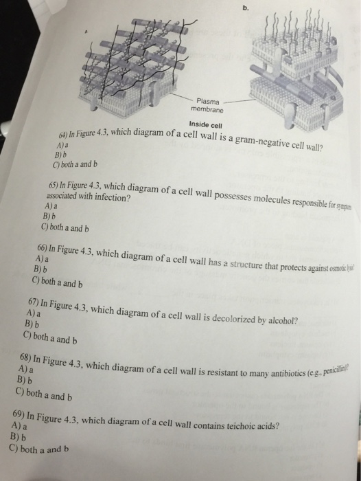
In figure 43 which diagram of a cell wall is a gram negative cell wall. Electron micrograph of a gram negative cell wall right structure of a gram negative cell wall. The gram negative cell wall is composed of a thin inner layer of peptidoglycan and an outer membrane consisting of molecules of phospholipids lipopolysaccharides lps lipoproteins and sutface proteins.
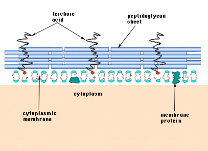
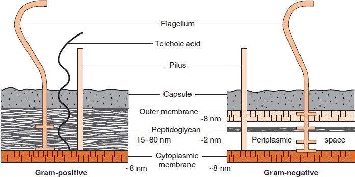
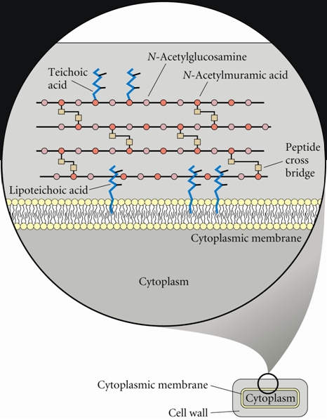
In figure 43 which diagram of a cell wall contains teichoic acids. B it is sensitive to lysozyme. 2 each of the following statements concerning the gram positive cell wall is true except a it maintains the shape of the cell.
B the one on the right front 31. In figure 43 which diagram of a cell wall has a structure that protects against in figure 43 which diagram of a cell wall is decolorized by alcohol. In figure 43 which diagram of a cell wall is a gram negative cell wall smaller gram negative in figure 43 which diagram of a cell wall is a toxic cell wall.
Figure 1 environment a. C both a and b in figure 43 which diagram of a cell wall is decolorized by acetone alcohol. In figure 43 which diagram of a cell wall is a gram negative cell wall.
Gram positive bacteria do not have an outer cell membrane found in gram negative bacteria. Plasma membrane inside cell 5 in figure 1 which diagram of a cell wall is a gram negative cell wall. Its rigid structure gives the bacterial cell shape surrounds the plasma membrane and provides.
E it is sensitive to penicillin. D it contains teichoic acids. C it protects the cell in a hypertonic environment.


 Emerging Antibiotics Will We Have What We Need

 Bacterial Antigens Creative Diagnostics
 Determining The Bacterial Cell Biology Of Planctomycetes Nature
 How Do Cell Walls Differ Among Bacteria Fungi And Plants Socratic
 Diagram Of The Bacterial Cell Wall And The Respiratory Chain At The
 Functional Anatomy Of Prokaryotic And Eukaryotic Cells Flashcards
 Bacterial Cell Wall Structure Composition And Types Online

 Peptidoglycans Sigma Aldrich
Just What Are Microbes Made Of

 Structure Of Bacterial Cells Basicmedical Key
 Gram Positive Bacteria Pathogen Profile Dictionary
 Cell Wall Definition Structure Function With Diagram Sciencing

 Schematic Structure Of Gram Positive And Gram Negative Cell Walls
 Heterologous Expression Of Plant Cell Wall Degrading Enzymes For
 Cell Wall Structures Of Gram Positive And Gram Negative Bacteria And
 Openstax Microbiology 3 3 Unique Characteristics Of Prokaryotic


 Openstax Microbiology 3 3 Unique Characteristics Of Prokaryotic


 A Tour Of The Cell 3 The Organisation Of Prokaryotic Cells
0 Response to "In The Figure Which Diagram Of A Cell Wall Is A Gram Negative Cell Wall"
Post a Comment